Home
/ Hip Joint Muscles Diagram - Hip Joint Anatomy / As with all joints, a degree of cushioning is required to stop the two bones from grinding against each other.
Hip Joint Muscles Diagram - Hip Joint Anatomy / As with all joints, a degree of cushioning is required to stop the two bones from grinding against each other.
Hip Joint Muscles Diagram - Hip Joint Anatomy / As with all joints, a degree of cushioning is required to stop the two bones from grinding against each other.. Hip pain explained including structures anatomy of the hip and pelvis author juni 02, 2021. The four groups are the anterior group, the posterior group, adductor group. Diarthrodial joint with its inherent stability dictated primarily by its osseous components/articulations. The hip joint is made up of two bones: 5 facts about the anatomy of the pelvic cavity.learn vocabulary, terms and more with flashcards, games and other study tools.
The hip joint is a multiaxial joint and permits a wide range of motion; Yet the hip joint is also one of our most flexible joints and allows a greater range of motion than all other joints in the body except for the shoulder. The hip joint is made up of two bones: Hip joint (articulatio coxae) the hip joint is a ball and socket type of synovial joint that connects the pelvic girdle to the lower limb. These muscles can be grouped based upon their location and function.
Hip Strains Orthoinfo Aaos from orthoinfo.aaos.org Yet the hip joint is also one of our most flexible joints and allows a greater range of motion than all other joints in the body except for the shoulder. These muscles move the thigh toward the body's midline. In human anatomy, the muscles of the hip joint are those muscles that cause movement in the hip.most modern anatomists define 17 of these muscles, although some additional muscles may sometimes be considered. A locking, clicking or catching sensation in your hip joint. The hip joint is a ball and socket synovial joint, formed by an articulation between the pelvic acetabulum and the head of the femur. Start studying leg/ hip muscles. Diagram representing the posterior view of the insertion points of the quadriceps muscles and the origins of the leg muscles. Hip joint muscles diagram / updated 27 awesome core exercises for athletes to build.
Yet the hip joint is also one of our most flexible joints and allows a greater range of motion than all other joints in the body except for the shoulder.
Any injury or disease of the hip will adversely affect the joint's range of motion and ability to bear weight. Bones of the hip joint. In this joint, the head of the femur articulates with the acetabulum of the pelvic (hip) bone. Yet the hip joint is also one of our most flexible joints and allows a greater range of motion than all other joints in the body except for the shoulder. Start studying leg/ hip muscles. The hip joint, like the shoulder joint, is a multiaxial synovial joint that flexes, extends, adducts, abducts, medially rotates, and laterally rotates. They can be divided into three main groups: These muscles move the thigh toward the body's midline. 5 facts about the anatomy of the pelvic cavity.learn vocabulary, terms and more with flashcards, games and other study tools. Hip joint (articulatio coxae) the hip joint is a ball and socket type of synovial joint that connects the pelvic girdle to the lower limb. Learn vocabulary, terms, and more with flashcards, games, and other study tools. Smartdraw includes 1000s of professional healthcare and anatomy chart templates that you can modify and make your own. Laterally rotates the the thigh at the hip joint.
As with all joints, a degree of cushioning is required to stop the two bones from grinding against each other. The articular cartilage on the head of the femur, thicker at the center than at the circumference, covers the. The hip muscles encompass many muscles of the hip and thigh whose main function is to act on the thigh at the hip joint and stabilize the pelvis.without them, walking would be impossible. They can be divided into three main groups: The pubis, ischium, and ilium together constitute the pelvis while the thigh bone is the femur.
Hip Anatomy Hip Surgeon Columbia Sc Hip Treatment Charleston Sc from www.dumontortho.com Laterally rotates the the thigh at the hip joint. Ligaments are soft tissue structures that connect bones to bones.a joint capsule is a watertight sac that surrounds a joint.in the hip, the joint capsule is formed by a group of three strong ligaments that connect the femoral head to the acetabulum. These are often divided into four groups according to their orientation around the hip joint: The bones of the hip include the femur, the ilium, the ischium, and the pubis. Press the back of your left knee into. Hip pain can sometimes be caused by diseases and conditions in other areas of your body, such as your lower back. In this joint, the head of the femur articulates with the acetabulum of the pelvic (hip) bone. Think of a model walking down the runway swinging her hips side to side.
A locking, clicking or catching sensation in your hip joint.
The bones together make up the hip. Learn vocabulary, terms, and more with flashcards, games, and other study tools. Yet the hip joint is also one of our most flexible joints and allows a greater range of motion than all other joints in the body except for the shoulder. Snapping hip syndrome is the result of the iliopsoas tendon subluxing over the greater trochanter or ilopectineal eminence. Many hip labral tears cause no signs or symptoms. In this type of joint, the working ends of the bones are surrounded by a membrane and inside this membrane is a thick fluid called synovial fluid. Several groin muscles are attached to the femur bone in the. The hip muscles encompass many muscles of the hip and thigh whose main function is to act on the thigh at the hip joint and stabilize the pelvis.without them, walking would be impossible. The hip joint is one of the most flexible joints in the entire human body. Ligaments are soft tissue structures that connect bones to bones.a joint capsule is a watertight sac that surrounds a joint.in the hip, the joint capsule is formed by a group of three strong ligaments that connect the femoral head to the acetabulum. The thighbone or femur and the pelvis join to form the hip joint. Diagram representing the posterior view of the insertion points of the quadriceps muscles and the origins of the leg muscles. Diarthrodial joint with its inherent stability dictated primarily by its osseous components/articulations.
The hip joint is an intricate structure including hip bones, hip articular cartilage, muscles, ligaments and tendons, and synovial fluid. The thighbone or femur and the pelvis join to form the hip joint. In this joint, the head of the femur articulates with the acetabulum of the pelvic (hip) bone. In this type of joint, the working ends of the bones are surrounded by a membrane and inside this membrane is a thick fluid called synovial fluid. Hip flexion is maximal with a high, forward kick that brings the leg above the level of the waist.
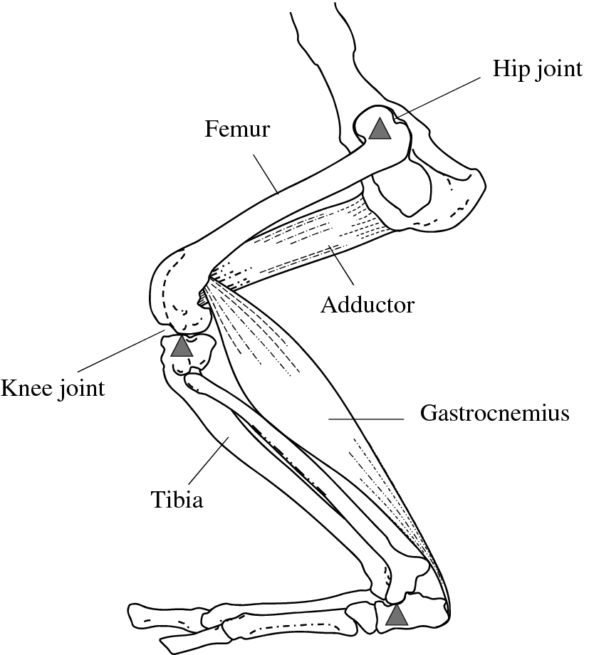
Figure 1 Morphological Differences Of Hindlimb Levers Between Wild And Farmed American Mink Neovison Vison And Implications For Reintroduction Of Mustelids Springerlink from media.springernature.com It allows us to walk, run, and jump. These are often divided into four groups according to their orientation around the hip joint: The movement at the joint depends on the anatomy of the joint and its axes of movement. Several groin muscles are attached to the femur bone in the. The ball is the rounded end of the. These muscles move the thigh toward the body's midline. Ligaments, tendons, and muscles play an important role in the function of the hip. The hip joint is a multiaxial joint and permits a wide range of motion;
The hip joint is an intricate structure including hip bones, hip articular cartilage, muscles, ligaments and tendons, and synovial fluid.
Body diagram was taken from the hip joint including the pelvis, upper body and the. Lie on your back and pull your right leg into your chest. Bones of the hip joint. Hip muscles anatomy hip anatomy human body anatomy muscle anatomy leg muscles diagram muscle diagram lower. Leg hip muscles diagram / physical therapy guide to groin strain choosept com. Flexion, extension, abduction, adduction, external rotation, internal rotation and. 5 facts about the anatomy of the pelvic cavity.learn vocabulary, terms and more with flashcards, games and other study tools. It allows us to walk, run, and jump. Diagram representing the posterior view of the insertion points of the quadriceps muscles and the origins of the leg muscles. Start studying leg/ hip muscles. Hip pain explained including structures anatomy of the hip and pelvis author juni 02, 2021. In human anatomy, the muscles of the hip joint are those muscles that cause movement in the hip.most modern anatomists define 17 of these muscles, although some additional muscles may sometimes be considered. Hip pain on the outside of your hip, upper thigh or outer buttock is usually caused by problems with muscles, ligaments, tendons and other soft tissues that surround your hip joint.
Leg hip muscles diagram / physical therapy guide to groin strain choosept com hip muscles diagram. Hip muscles anatomy hip anatomy human body anatomy muscle anatomy leg muscles diagram muscle diagram lower.